Submandibular Gland Cancer
The submandibular gland is the second largest salivary gland, where salivary gland tumors are the second most common. Half of the submandibular gland tumors are benign and half are malignant. The most common benign submandibular gland tumor is pleomorphic adenoma. The most common malignant submandibular salivary gland tumors (submandibular gland cancer) are adenoid cystic carcinoma and mucoepidermoid carcinoma.
The causes of submandibular gland tumors are unknown. Risk factors such as exposure to radiation, exposure to chemicals, tobacco use, and alcohol consumption may have an impact on the development of submandibular salivary gland cancer. Submandibular gland tumors may give late signs and symptoms. Avoiding risk factors may be beneficial in submandibular gland cancer prevention.
The diagnosis of a submandibular gland tumor can be made by biopsy. Survival rate may vary depending on the stage, spread, type of submandibular gland cancer, and the age of the patient. The main treatment for a submandibular gland tumor is surgery. Where necessary, the treatment also includes radiation therapy and sometimes chemotherapy.
The Submandibular Gland
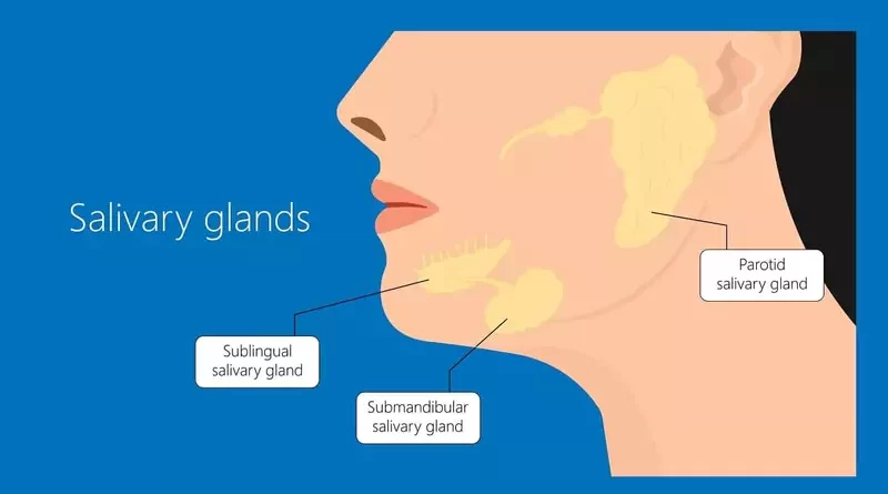
We have 3 pairs of major salivary glands. These are the parotid, submandibular, and sublingual salivary glands. Apart from these, there are also many minor salivary glands. The submandibular gland is the second largest salivary gland after the parotid gland. But despite this, the submandibular gland secretes the most saliva.
One pair of submandibular glands is located on both sides under the jawbone. The saliva-carrying submandibular gland duct (Wharton’s duct) opens under the tongue, just behind the lower anterior incisor teeth, to the sides of the midline.
The marginal mandibular branch of the facial nerve that moves the lower lip muscles passes through the surface of the sheath of the submandibular gland. During surgery, it may not always be easy to recognize this thin nerve.
The second most common site of salivary gland tumors is the submandibular gland. In general, as the size of the salivary gland decreases, the risk of malignancy of the salivary gland tumor increases. Clinically, most parotid tumors are benign but half of the submandibular gland tumors are benign and half are malignant.
Signs and Symptoms
A benign tumor of the submandibular gland is a slowly (over months and years) growing mass under the chin. Benign submandibular gland tumors can turn into malignant tumors over time. Malignant tumors of the submandibular gland develop and grow in a shorter period of time (within weeks or months). Submandibular gland tumors rarely occur in both glands at the same time.
Salivary stones (sialolithiasis) obstructing the duct of the submandibular gland are more common than its tumors. Sialolithiasis is a common disease of the salivary glands and occurs mostly in the submandibular gland. This condition can lead to occasional swelling, redness, and submandibular gland pain that heals later. In addition, when a growing sublingual gland tumor exerts pressure on the Wharton’s duct, obstructive swelling and/or sialadenitis of the submandibular gland may develop.
Salivary stones can sometimes cause inflammation of the submandibular gland (sialadenitis). In case of inflammation in the gland, swelling, pain, and redness increase more. However, antibiotic treatment or the movement of the stone over time provides recovery in the gland. However, the swelling due to the tumor does not regress and does not shrink, it progresses further over time.
Lump under the jawbone on one side
The tumor of the submandibular gland, whether benign or malignant, manifests itself as swelling, lump, mass, or growth under the chin, usually present for more than 2-3 weeks.
In benign tumors, this lump is a medium-hard, rubbery, mobile submandibular mass between two fingers.
In malignant submandibular gland tumors, the hardness of the mass is higher than in benign tumors. It is more difficult to move the mass between the fingers. If the mass is attached to the muscle or jawbone, it is more immobile.
Subsequent jaw pain on one side
Pain occurs as submandibular gland cancer develop and invade surrounding tissues (such as nerves). This one-sided submandibular gland pain that starts later turns into persistent jaw pain. As benign submandibular gland tumors do not invade surrounding tissues, they generally have a painless course.
Paralysis of the lower half of one side of the face
If submandibular gland cancer invade the marginal mandibular branch of the facial nerve that moves the lower lip, there may be numbness, tingling, weakness, or even paralysis in that area. In this respect, weakness or paralysis of the lower half on one side of the face may be a sign of a submandibular gland cancer. Such findings generally do not occur in benign tumors of the submandibular gland.
Swollen lymph nodes in the neck on one side
There is a possibility that malignant tumors of the submandibular gland spread to the lymph nodes in the neck. In this case, swelling may occur on that side of the neck. Benign tumors of the submandibular gland do not cause signs such as swelling and enlargement in the lymph nodes in the neck.
Tethering or ulceration of overlying skin
If the submandibular gland cancer are close to the skin, they may cause skin changes, redness, or ulcer formation due to skin involvement over time. Benign submandibular gland tumors do not cause redness, tightness, or other changes in the overlying skin.
Without any symptoms
Submandibular salivary gland tumors may give late signs and symptoms. Especially malignant submandibular salivary gland tumors, when they give signs and symptoms, they can be in the late stages. Sometimes, imaging tests taken for other reasons detect them incidentally.
Causes
Tobacco use, radiation exposure, occupational chemical exposures, and sometimes alcohol consumption are blamed for the occurrence of submandibular gland cancers. But there is no clear scientific evidence. Viruses, diet, or other factors have not been proven either. The causes of benign tumors of the submandibular gland are even more uncertain.
Risk Factors
For salivary gland tumors, including submandibular glands, the gender distribution is equal, and most cases occur in the sixth decade. Submandibular gland tumors can be seen at any age, including childhood. However, benign submandibular gland tumors are more common in the young and middle age group, and malignant tumors are more common in elderly people.
Being older
Although submandibular salivary gland tumors can occur at any age, malignant tumors are most common in older adults.
Tobacco use
Tobacco use has an effect on the formation of many head and neck cancers. In this respect, malignant submandibular gland tumors may also be associated with tobacco use. Because tobacco use is common in many patients. Apart from this, tobacco use is also a risk factor in the transformation of benign submandibular gland tumors into malignant tumors.
Exposure to radiation
Exposure of the head and neck region to radiation during radiation therapy or in any case is a risk factor. Nowadays, suspicions are increasing due to the electromagnetic radiation emitted by mobile phones.
Exposure to chemicals
People who work with certain substances known to be carcinogenic may have an increased risk of developing submandibular salivary gland cancer. In this respect, regular occupational exposures are important. The chemical industry, mining, plumbing, rubber, or nickel industries are among the risky jobs in terms of submandibular gland cancers.
Alcohol consumption
Unlike some head and neck cancers associated with alcohol consumption, the role of alcohol in salivary gland tumors is unclear. There is no definite information about the effect of alcohol consumption on the development of submandibular salivary gland cancers.
Types of Tumors
Tumors are masses formed by the uncontrolled proliferation of cells. There are tumors of the salivary glands as well as tumors of any tissue in the body. Tumors are generally benign or malignant. Half of the submandibular salivary gland tumors are benign and half are malignant. The malignant submandibular gland tumor is often called submandibular gland cancer.
Benign submandibular gland tumors
Benign submandibular gland tumors grow slowly. They grow by pushing the surrounding tissues and growth occurs over months or even years. Benign submandibular gland tumors usually have capsules. They do not spread to the surrounding tissues and lymph nodes. They do not metastasize to organs in the distant region. The most common benign submandibular gland tumors are the following;
Pleomorphic adenoma (benign mixed tumor)
Pleomorphic adenoma is the most common benign tumor of both the salivary glands as well as the submandibular gland. Normally, pleomorphic adenomas grow slowly. However, they can also turn into malignant tumors over the years. In this case, the mass under the jaw, which grows very slowly, begins to grow rapidly. Scientific studies have shown that one-tenth of submandibular pleomorphic adenomas that have existed for over fifteen years can become cancerous.
Basal cell adenoma
Basal cell adenoma is a benign tumor that can develop from the submandibular gland, although it mostly arises from the parotid gland.
Oncocytoma
Oncocytoma is a benign tumor that mostly originates from the parotid gland. However, it can also develop from the submandibular salivary gland. Oncocytoma originates from a type of salivary gland cell called oncocyte.
Other benign tumors
In rare cases, benign tumors such as myoepithelioma, canalicular adenoma, sialoadenoma papilliferum may develop from the submandibular salivary gland. Apart from this, submandibular mass originating from vessels such as lymphangioma (cystic hygroma), hemangioma, and salivary gland cysts may also occur in the submandibular gland.
Submandibular gland cancers
Malignant tumors of the submandibular gland are usually not encapsulated. They grow fast and spread to regional lymph nodes. They invade surrounding tissues (eg nerves) and develop by disrupting their functions. Malignant submandibular gland tumors can metastasize to distant areas such as the lungs, liver, brain, and bones. The most common submandibular gland cancers are the following;
Adenoid cystic carcinoma
Adenoid cystic carcinoma is the most common malignant tumor of the submandibular gland. This tumor penetrates the nerve fibers in the area and uses them as a way to spread. Adenoid cystic carcinoma spreads through the blood rather than lymph.
Adenoid cystic carcinomas tend to return years after treatment, and lung metastases are common. However, adenoid cystic carcinoma is a slower-growing tumor with a long history of the disease.
Adenoid cystic carcinoma may show little change over a long period of time. But, then suddenly spreads extensively to surrounding tissues. These tumors are more common in people of middle age and older.
Mucoepidermoid carcinoma
Mucoepidermoid carcinoma is the most common salivary gland cancer and also one of the most common cancers of the submandibular salivary gland. Although it is a relatively slow-growing type, it is known to behave aggressively when mucoepidermoid carcinoma occurs in the submandibular gland. The patients are generally middle-aged and older adults. However, younger people can also get mucoepidermoid carcinoma of the submandibular gland.
Malignant mixed tumors
Malignant mixed tumors are submandibular salivary gland cancers specific to older ages. Carcinoma ex pleomorphic adenoma is the most common of these. This tumor is the untreated pleomorphic adenoma that has turned into a malignant tumor over many years. In this case, the mass under the chin that has existed for years suddenly begins to grow rapidly. It is a highly aggressive tumor with very low cure rates regardless of treatment.
Acinic cell carcinoma
Acinic cell carcinoma, a type of adenocarcinoma, accounts for about one-tenth of malignant tumors of the salivary glands. Although this tumor mostly develops from the parotid gland, it is also seen in the submandibular gland. However, acinic cell carcinomas are tumors with low malignant potential and grow slowly.
Although acinic cell carcinomas are malignant, they are encapsulated tumors and are unlikely to spread to distant organs. These tumors, which are mostly seen in young adults, have a more benign course compared to other submandibular salivary gland cancers. Acinic cell carcinoma can also be seen in children and elderly people.
Adenocarcinoma
Many types of adenocarcinomas occur in the submandibular gland. Some of these tend to be slow-growing (eg acinic cell carcinoma) and generally have a good prognosis. But types such as salivary duct carcinoma and oncocytic carcinoma are more likely to be aggressive and have a less positive outlook
Other cancers
Sometimes, non-Hodkin lymphoma, squamous cell carcinoma, and other cancers can also develop from the salivary glands. Of these, squamous cell carcinomas are mainly seen in elderly people. These cancers can develop after radiation therapy and tend to have a poorer outlook.
Although rare, Non-Hodgkin lymphomas can develop from immune system cells in the submandibular salivary glands. In this case, they affect people with Sjögren’s syndrome. Non-Hodgkin lymphomas are tumors that can be found in both submandibular glands at the same time.
Diagnosis
A patient who suspects submandibular salivary gland cancer should first consult an otolaryngologist (ear, nose, and throat doctor). The diagnosis process begins when the patient notices a swelling and mass under the jaw and consults a doctor. The doctor first listens to the patient’s complaints. Then the doctor asks the patient various questions. The doctor then proceeds to the examination phase.
Physical exam
The otolaryngologist makes a complete head and neck examination of the patient. The doctor carefully examines the tumor under the chin in terms of size, hardness, mobility, adhesion to surrounding tissues. Then the doctor checks for reduced movement or weakness in the muscles of the lower half of the face.
Imaging tests
The doctor who suspects a submandibular salivary gland tumor on examination usually requires one or more of the tests such as ultrasonography (USG), magnetic resonance imaging (MRI), or computed tomography (CT). If the tumor has no relationship with the oral cavity or throat, imaging tests are first performed before tissue sampling. The radiologist evaluates the image of the submandibular mass in imaging tests and reports it to the surgeon.
Ultrasound imaging is the preferred tool for the initial assessment of tumors in the submandibular gland. Ultrasound imaging of the superficial structures is excellent and it does not carry any risk of radiation. Apart from this, ultrasonography is often the first imaging test used because it is the easiest to reach.
Magnetic resonance imaging provides more information about soft tissues and their properties. An MRI with an intravenous drug (contrast agent) has become an important imaging test to examine a salivary gland tumor.
Benign tumors of the submandibular salivary gland are well-circumscribed on MRI or CT and do not occupy the surrounding tissues. Malignant tumors, on the other hand, often appear in a way that invades the surrounding tissues depending on their location and the borders cannot be clearly identified.
In cases where adenoid cystic carcinoma, which has the risk of spreading to distant organs such as the lungs, is diagnosed in the submandibular gland, examinations such as positron emission tomography (PET) or lung CT are required to investigate the presence of metastasis.
Removing a tissue sample
It is necessary to take tissue samples from the submandibular mass to confirm the diagnosis. The doctor takes tissue samples from tumors that are not associated with the oral cavity or throat and are in the form of swelling under the skin, with a fine needle biopsy.
Unless the tumor reaches the skin surface, it is not safe to take tissue samples by cutting the skin. Otherwise, the tumor may spread and become more difficult to treat. Apart from this, there is a possibility of damage to the branch of the facial nerve under the chin. In this respect, the doctor takes a biopsy from the submandibular gland tumor with a needle.
Fine needle biopsy is a safe method that does not cause the tumor to spread. The doctor often uses ultrasound to guide the needle during the biopsy. Needle biopsy does not cause much physical discomfort to the patient, and after this procedure, the patient can return to his daily life.
The surgeon can skip the fine-needle biopsy stage, which provides a lot of information about the type of submandibular gland tumor. Instead, the surgeon surgically removes the entire tumor and sends it to the pathologist for pathological examination. The pathologist reaches the final diagnosis by examining the removed tumor. This method leads the pathologist to a more precise diagnosis. That is why the diagnosis made by fine needle biopsy can change with the pathologist’s examination of the removed tumor.
Treatment
The main treatment of all submandibular salivary gland tumors, regardless of whether they are benign or malign, is the surgical removal of the tumor. Because even if the submandibular gland tumor is benign, it will continue to grow over time. Apart from this, there is a possibility that a benign submandibular gland tumor can turn into a malignant tumor over time. All of these can make the tumor difficult to treat. In addition, there are transitional zone tumors of the submandibular gland, which the pathologist may not be able to determine whether they are benign or malignant in nature.
If the pathological examination of the tumor of the submandibular gland completely removed shows that it is a benign tumor, there will be no need for additional treatment (radiation therapy, chemotherapy). Because surgical treatment is usually sufficient for benign tumors. In addition, radiation therapy and chemotherapy have no place in the treatment of benign tumors.
After complete removal of submandibular gland tumors with low malignancy potential, additional treatments such as radiation therapy and chemotherapy may not be required. However, submandibular gland cancers with high malignancy potential require additional treatments after surgery. If the patient’s health condition is not suitable for surgery, or if the submandibular gland cancer has spread to the extent that surgery will not work, other treatment options without surgery may come forward.
Treating doctors
Problems related to the submandibular salivary gland in the lower jaw and neck area mainly concern otolaryngologists. Otolaryngologists, experienced in head and neck surgery perform submandibular salivary gland tumor surgeries. Radiologists, radiation oncologists, medical oncologists, plastic surgeons, neurosurgeons, dentists, psychologists can also participate in the diagnosis and treatment process depending on the situation. Therefore, in the presence of a mass under the chin, it is the best choice to consult an experienced otolaryngologist.
Surgery
Submandibular gland tumor surgery is performed under general anesthesia with an incision under the chin. However, the surgeon may perform the surgery via an intraoral approach, depending on the location of the tumor in the salivary gland.
The marginal mandibular branch of the facial nerve that moves the lower lip muscles passes through the surface of the sheath of the submandibular gland. Apart from this, the hypoglossal nerve that moves the tongue passes through the lower deep part of the submandibular gland. The lingual nerve, which provides the tongue’s sense of taste and touch, passes through the upper deep part of the gland. It is very important to recognize and protect these nerves during surgery.
A special device – the nerve monitor makes it easy for the surgeon to recognize and protect nerves during surgery. However, botox, which is a very common cosmetic application today, may prevent the nerve monitor device from functioning during surgery. If the patient has had botox within the last 6 months, the surgeon must know this. Because the effect of botox can last up to 6 months.
Tumor removal
Surgery for the submandibular gland tumors will vary depending on the location of the tumor in the salivary gland. The nerve that passes through the surface of the sheath of the submandibular gland and moves the lower lips is very thin. It is not always easy to recognize this nerve during tumor removal. In this case, the nerve monitor device facilitates the recognition and protection of this nerve branch.
Usually, the surgeon removes the submandibular gland completely with the tumor. To protect the branch of the facial nerve, gland removal surgery should be performed under the capsule of the gland, leaving the capsule in place. However, cases, where a malignant tumor invades the nerve or adhesions as a result of chronic inflammation, can make this difficult. Tongue nerves such as hypoglossal and lingual are relatively easy to protect because they are thicker than the nerve of the lower lip
Lymph node removal
In surgeries for submandibular gland removal, the neck lymph nodes with the highest probability of the malignant tumor spreading are removed (neck dissection). This procedure has become a standard treatment for many malignant tumors located in the face, head, and neck regions.
The surgeon performs the neck dissection in cases of lymph node metastasis, usually detected by examination or imaging tests in the neck. However, the surgeon can also perform this procedure to prevent future metastases, even when there is no visible metastasis.
If the pathologist diagnosed submandibular gland cancer as a result of the pathological examination of the tumor after surgery, the surgeon performs a second surgery to remove the neck lymph nodes on the same side of the tumor. Submandibular tumors with a low malignancy potential may not require neck dissection. In benign tumors of the submandibular gland, there is no need for neck dissection surgery.
Reconstruction
After removing the submandibular gland tumor (usually with the gland), the surgeon goes into the repair phase. During surgery, the surgeon may have removed skin, muscle, nerves, or bone along with the tumor. For cosmetic and functional repair of the face, mouth, tongue, throat, or jaw, the surgeon can make skin, muscle, nerve, or bone tissue transfers from other parts of the body.
Normalization after surgery
After submandibular gland tumor surgery, the patient usually stays in the hospital for several days. A tube (drain) placed to expel blood and fluid accumulation from the surgical wound site is removed after a day or two. However, the bandage remains for a few more days to prevent blood and fluid accumulation in the surgical area.
After submandibular gland tumor surgery, the patient takes pain relievers for the first few days. These are often enough to cope with the pain. To prevent infection, the patient also takes antibiotics for a few days.
After surgery, there may be swelling in the face and neck as a result of blood and fluid accumulation. Apart from that, postoperative bandages can also cause pain and swelling. Such situations pass over time
Radiation therapy
The radiation oncologist applies the radiation produced by special devices to the tumor area and the areas where the tumor is likely to spread. Although the main treatment of submandibular salivary gland cancers is surgery, radiation therapy is also added to the treatment to increase the effectiveness of the treatment. Radiation therapy increases the patient’s chance of getting rid of cancer. However, radiation therapy is not the primary and sole treatment for submandibular salivary gland cancers.
The patient receives radiation therapy after surgery if the submandibular gland tumor has a high malignancy potential. If the submandibular gland tumor has a low malignancy potential and the surgeon has removed the tumor completely, the patient may not receive radiation therapy. Sometimes, some persistent benign tumors of the submandibular gland may also require radiation therapy.
Chemotherapy
Chemotherapy uses drugs, which seek out and kill cancer cells in the body. It is not a standard treatment for submandibular salivary gland tumors. Even in cases where tumors of the submandibular gland are malignant, chemotherapy as additional therapy is rarely required. Treatment of the submandibular gland tumors with medication alone will not work unless there is an inflammation-like condition.
Chemotherapy is essential in the treatment of lymphoma of the salivary gland. Apart from this, in the presence of metastasis and in submandibular gland cancers that cannot be completely removed or recurred, the patient receives chemotherapy.
Targeted therapy
So far, targeted therapy drugs have not been seriously effective in submandibular salivary gland cancers. Targeted therapy research is still at an early stage in the treatment of salivary gland cancers, including the submandibular gland.
Nerve functions after surgery
Protection of the lower lip and tongue nerves is an important part of submandibular gland tumor surgery. In cases where the submandibular salivary gland tumor affects these nerves, the patient feels weakness in the lower lip or tongue. This weakness may manifest itself in the form of asymmetry in the lower lip, usually while smiling, or difficulty in moving the tongue. If the tumor has little effect on the nerves, these problems can usually resolve within weeks after surgery.
If submandibular gland cancer has infiltrated the lower lip nerve and tongue nerves, the surgeon can remove these nerves along with the tumor. In these patients, asymmetry, weakness, and paralysis are observed to a certain extent in the lower lip and/or tongue before the surgery. The surgeon repairs the damaged nerves with a nerve graft taken from another part of the body. However, even in experienced hands, these nerves rarely fully recover functionally.
Salivary functions after surgery
Removing one of the three pairs of large and many minor salivary glands that secrete saliva through various ducts into the mouth does not cause a decrease in the amount of saliva or dry mouth. Even though the submandibular gland is the most saliva-producing, other submandibular glands and other glands take over the function of the removed gland. However, the patient, who also received radiation therapy after surgery, may experience dry mouth for a while. ADA approved mouth moisturizers (eg, Biotene Dry Mouth Oral Rinse, or as a different choice: TheraBreath Dry Mouth Oral Rinse, etc.) and other relevant oral care products may be beneficial for such patients.
Appearance after surgery
The surgeon performs the submandibular gland tumor surgery with an incision under the chin under aesthetic principles. However, in dark-skinned people, the surgical incision can heal with a distinct pattern. This situation is actually more related to the genetic characteristics of the person
To reduce the possibility of significant scar healing of the surgical incision, patients should not show the incision area to the sun for up to a year. Patients should protect the area with high protection factor creams or clothing accessories. Apart from this, some medicines in the form of cream and gel can make the scar in the chin area less prominent.
In terms of appearance, the patient does not feel the absence of the submandibular salivary gland removed with the tumor. Surgical removal of large tumors can create a dimple in the lower jaw area. However, with the increase of adipose tissue over time and also with gravity, the dimple under the chin may disappear.
Speaking and feeding after surgery
After the surgery, the patient is not given liquid or food orally for a period of time until the effect of anesthesia disappears. After a while, the patient starts feeding with soft, liquid foods. The patient starts to eat normal foods in time. However, after extensive surgeries performed for large tumors under the chin, it may take longer for the patient to return to a normal diet. Unless some muscles, nerves, jawbone, or neck skin are removed due to submandibular gland cancer, functions such as tongue and lip movements and speech are not permanently affected.
The secretion formed by the salivary glands helps digestion by keeping the mouth and throat moist. In addition, saliva creates a defense mechanism against microorganisms that cause infection in the mouth and throat. After a while, the side effects of radiation therapy, which causes a decrease in saliva secretion, may cause wounds in the patient’s skin, mouth, and throat areas. However, after these effects pass over time, the patient’s saliva and swallowing problems also pass.
Prognosis
If submandibular gland cancer is found early and treated, the prognosis is good. The prognosis of salivary gland cancers is better in children and adolescents because of the lower frequency of metastasis, less local soft tissue spread, and relatively different types of cancer. Overall survival is better for children than for adults after successful surgical treatment.
Regardless of the type of malignant tumor of the submandibular gland, adverse prognostic factors in survival are older age, advanced stage of cancer, presence of distant metastasis, presence of pain, nerve involvement, and comorbidities. Distant metastases most commonly occur to the lungs, bone, liver, and brain. Distant metastases are the main cause of death in submandibular gland cancers.
Follow-up care
The main goals of regular doctor check-ups after cancer treatment are to detect recurrence and manage any treatment complications that may occur. Long-term follow-up is very important for all salivary gland tumors, including the submandibular gland. Most recurrences occur in the first 3 years after treatment, except for tumors with log malignancy potential and adenoid cystic carcinoma.
Patients should receive regular follow-up within 5 years after treatment. Then, regular follow-up should continue every 1 year.
All salivary gland tumors require observation for up to 20 years after treatment. Adenoid cystic carcinoma should be approached more carefully, especially because of delayed recurrence or tendency to metastasis. Annual chest imaging is important for submandibular tumors with high malignancy potential because of the risk of frequent lung metastasis. Patients who receive neck radiation should have their thyroid hormone levels checked several times a year.
Recurrence of cancer
Submandibular gland tumors can recur after treatment. In these cases, if possible, the surgeon attempts to remove the tumor completely along with the surrounding tissues by repeat surgery. Repeat surgeries have some risks. Because it will be more difficult to recognize and protect the lower lip and tongue nerves in the region due to the healing tissues caused by the previous surgery.
Prevention
The cause of malignant tumors of the submandibular salivary gland is unknown. The causes of benign submandibular gland tumors are much more uncertain. In this respect, the ways to prevent submandibular gland cancers, the exact cause of which is not known, are also unclear. But it can be helpful to avoid some risky things.
Reduce radiation exposure
In terms of submandibular gland tumors, even if it is for treatment, the radiation received in the past under the jaw (especially during dental treatments) or to the neck region is a risk factor. Especially if the person is elderly, the negative effect of this radiation is more.
In this respect, it is important to avoid radiation exposure to the under-chin and neck area as much as possible. In addition, in terms of close exposure to electromagnetic radiation, using the mobile phone as little as possible or using it with a headset may reduce the risk.
Reduce chemical exposure
People who work with certain substances known to be carcinogenic due to their profession should work in a more protected environment as much as possible. These people mostly work in the chemical industry, mining, plumbing, rubber, or nickel industries
Quit smoking
Tobacco use may be a cause of submandibular gland cancers, as it exposes the person to carcinogenic substances. Especially if the person is older, this risk will increase even more. Not using tobacco can reduce the risk of many head and neck cancers.
Reduce alcohol consumption
Alcohol consumption is an important risk factor for various head and neck cancers. However, the role of alcohol in the development of submandibular gland cancers is uncertain. Limiting alcohol consumption may be beneficial in reducing the risk. Even the use of alcohol-containing oral care products is risky. Because they regularly and continuously expose the mouth to alcohol. In this respect, the use of alcohol-free products (eg, ADA approved mouthwash: TheraBreath Fresh Breath, CloSYS Sensitive Gentle Mint, CloSYS Ultra Sensitive Unflavored, or as a different choice: Tom’s of Maine Natural Wicked Fresh, etc.) is important.
Live a healthy life
A healthy diet, good immunity, and high morale are important to reduce the risk of developing all types of cancer, including submandibular gland cancer. Some studies have shown that a diet rich in vitamin C and low in cholesterol can be effective in preventing salivary gland cancer.
References
American Cancer Society: “What’s New in Salivary Gland Cancer Research and Treatment?”
Cedars-Sinai: Health Library: “Salivary Gland Disease and Tumors”
Mayo Clinic: Diseases and Conditions, “Salivary gland tumors”
National Cancer Institute:“Salivary Gland Cancer Treatment”
NCCN Clinical Practice Guidelines in Oncology: “Head and Neck Cancers, Version 2.2020”
PubMed: “Tumors of the submandibular gland: clinicopathologic analysis of 23 patients”